Computer Vision in Pharmaceutical R&D: Why Microscopy Still Requires Custom AI
Computer vision is already well established across pharmaceutical R&D and manufacturing. It supports applications ranging from automated visual inspection and quality control to digital pathology, laboratory automation and microscopy image analysis. As AI models and commercial image analysis platforms continue to improve, it’s fair to ask whether custom computer vision is still necessary.
From our experience, the answer depends on the problem you’re trying to solve.
For standardized image analysis tasks, commercial software often provides everything researchers need. But microscopy projects in pharmaceutical R&D are rarely standardized. They involve proprietary materials, specialized imaging techniques and scientific questions that require more than detecting or classifying structures in an image.
We’ve seen this firsthand while developing a computer vision solution for scanning electron microscopy (SEM)-based material analysis. The project reinforced something we’ve observed across scientific imaging: the real challenge isn’t applying AI to microscopy. It’s designing analysis workflows that produce reliable, objective measurements researchers can act on with confidence. We bring that same discipline from years of building computer vision systems in environments where measurement accuracy matters, and where unreliable outputs can have real consequences.
In this article, we’ll explore why microscopy remains one of the most demanding computer vision applications in pharmaceutical research, where commercial tools work well and where custom AI continues to create value.
Computer Vision Already Supports the Entire Pharmaceutical Lifecycle
Computer vision is no longer an emerging technology in this domain. Many applications of computer vision in pharmaceutical technology are already well established across both research and manufacturing.
In production environments, AI-powered vision systems commonly perform visual inspection of products and packaging, verify labels, inspect fill levels, and detect defects.
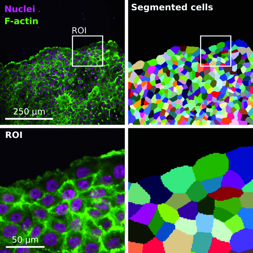
Research and development presents a distinct set of challenges. Researchers analyze visual data generated through techniques such as microscopy, spectroscopy, laboratory imaging, and other scientific methods. They use these images to characterize biomaterials and other particles and to inspect cells and tissues. This analysis supports applications such as digital pathology, laboratory automation, and high-content screening.
Although these use cases all fall under computer vision, they rarely require the same solution. Every dataset is different, every research question is different, and the information researchers need to extract depends entirely on the scientific objective.
Why Microscopy Is Different
Identifying cells in microscopy images is fundamentally different from traditional visual inspection.
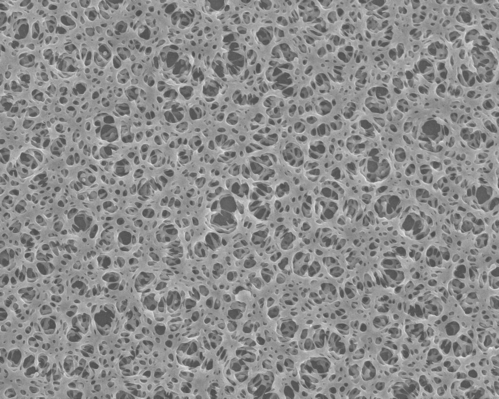
Unlike conventional computer vision datasets, microscopic images frequently contain irregular structures, overlapping features, highly detailed textures and objects that exist across multiple focal planes. Depending on the imaging technique, even small differences in illumination or sample preparation can significantly affect the appearance of the material that we’re analysing.
In microstructural characterization and analysis specifically, the objective is not simply to classify images, but to quantify structural characteristics, develop new metrics and discover the relationship between microstructure properties that support scientific conclusions.
In our work, one thing we’ve consistently observed is that developing the model is rarely the hardest part of the project.
The more difficult challenge is deciding what should actually be measured.
Researchers and lab technicians can often identify meaningful differences between samples immediately because of years of domain expertise. Translating that intuition into quantitative metrics that can be extracted consistently from thousands of images requires close collaboration between experts and AI engineers. Before training begins, both teams need to agree on which structural characteristics are scientifically relevant and how those characteristics should be represented in the data.
Commercial Software Solves Many Problems, but Not Every Problem
Modern microscopy platforms already include increasingly sophisticated image analysis capabilities. For standardized laboratory workflows such as object detection, segmentation, counting or basic morphological measurements, these solutions are often more than sufficient.
However, pharmaceutical research frequently extends beyond these routine applications. In areas such as drug delivery system optimization, researchers are increasingly using AI and machine learning to reduce experimental workload, accelerate formulation development and identify new design opportunities. As research questions become more specialized, image analysis must also evolve beyond standardized workflows.
The challenge arises when researchers need to answer questions that existing software was never designed to address. Pharmaceutical R&D often involves complex formulations, drug delivery systems and specialized imaging protocols, each with its own analytical objectives. Depending on the use case, researchers may need to characterize a formulation’s microstructure or analyze particle size and distribution. They may also investigate crystal morphology or study the structural properties of long-acting injectable (LAI) depots. Researchers can then examine how these characteristics relate to drug release and bioavailability. Commercial tools can extract general image features. However, they do not always provide the specific measurements needed to answer these questions.
Every Project: Different Question
Every project therefore asks a different scientific question. One team may need to quantify pore distribution or other microstructural properties. Another may investigate particle size or crystal morphology. A third may want to understand how microscopic characteristics relate to formulation or product performance. Although these problems can look similar from a computer vision perspective, they can require entirely different structures of interest, relevant measurements and analytical workflows.
In these situations, the challenge is not detecting structures. It is extracting the information researchers need to support scientific decision-making. This is where custom computer vision creates value. Rather than replacing commercial software, a custom model is designed around the client’s workflow, imaging data and research objectives, enabling the extraction of measurements that standard solutions cannot readily provide.
Beyond analytical capabilities, data governance is another important consideration in pharmaceutical R&D. Microscopy images and experimental data often represent valuable intellectual property and are subject to strict internal security policies. Companies can deploy custom CV solutions within their own infrastructure, allowing them to leverage AI while maintaining control over sensitive research data and complying with internal data governance requirements.
Lessons from Building a Microscopy Analysis Pipeline
One of our recent projects involved developing an AI-assisted workflow for analyzing scanning electron microscopy images. The workflow supported material characterization in an R&D environment. The material and manufacturing process remain confidential. However, many of the technical challenges are common to scientific imaging projects across the pharmaceutical and medical industries.
The project started with a familiar problem. Researchers could evaluate material performance at a high level. However, manually analyzing large collections of SEM images was slow and difficult to scale. Individual interpretation could also influence the results.
The goal was not to replace scientific expertise. Instead, the team aimed to create a more objective framework for consistently analyzing microscopic structures across much larger datasets.
The biggest technical challenge was annotation.
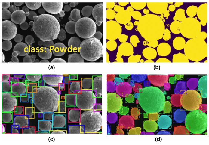
Unlike many computer vision applications with clearly defined object boundaries, SEM images contain irregular structures with complex textures and overlapping features. Researchers must invest substantial manual effort and collaborate closely with domain experts to create reliable pixel-level annotations. In some cases, researchers must also account for uncertainty in the annotation process during model development.
Another challenge involves deciding which characteristics to extract from the images.
Rather than producing segmentation masks as the final output, the goal was to convert microscopic structures into quantitative material metrics that researchers could analyse statistically. The workflow combined semantic segmentation, feature extraction, statistical analysis and visualization, creating a foundation for further material research rather than simply automating image processing.
Compared with manual evaluation, this approach provided faster analysis of large image collections, more consistent measurements and a scalable framework for correlating microscopic material characteristics with overall product performance.

AI Should Adapt to Research, Not the Other Way Around
One misconception about computer vision is that its primary purpose is automation.
In pharmaceutical R&D, we see it differently.
The real value is in making complex visual information measurable, reproducible and easier to interpret. Researchers still provide the scientific expertise and make the final decisions.
Computer vision contributes by performing image analysis consistently across hundreds or thousands of samples, allowing experts to spend more time interpreting results instead of performing repetitive measurements.
The most successful projects do not begin with selecting a neural network architecture or experimenting with the latest AI model. They begin by understanding the research question, evaluating the available imaging data and determining which measurements will generate meaningful scientific insight, as explained in our How We Work methodology.
Looking ahead
Computer vision will continue to play an increasingly important role across pharmaceutical research and manufacturing, but we believe its greatest value lies beyond automation.
As imaging technologies become more advanced, laboratories generate larger and more complex datasets than researchers can reasonably analyse manually. AI makes those datasets easier to process, but its real contribution is creating objective, reproducible measurements that strengthen scientific decision-making.
Commercial software will continue to solve many image analysis problems, and for standardized applications it is often the right choice.
However, when research moves beyond standardized workflows, custom computer vision can bridge the gap between complex microscopy images and the specific insights researchers need. The most successful projects begin not with selecting an AI model, but with understanding the scientific problem and designing image analysis around it.
Frequently Asked Questions
How is computer vision used in pharmaceutical research?
Computer vision can support many pharmaceutical R&D activities, including microscopy image analysis, biomaterial characterization, particle analysis, crystal morphology evaluation, laboratory automation and digital pathology. Its primary role is to convert complex visual information into objective, measurable data that supports scientific research.
Why is microscopy challenging for computer vision?
Microscopy images contain irregular structures, overlapping features, dense textures and variability introduced during sample preparation and imaging. These characteristics make annotation, segmentation and quantitative analysis considerably more challenging than conventional computer vision tasks.
Can commercial microscopy software solve every use case?
Commercial platforms are highly effective for many standardized laboratory workflows. However, research involving proprietary materials, specialized imaging techniques or unique scientific questions often requires custom computer vision models designed around the specific analytical objective.
When should pharmaceutical companies consider custom computer vision?
Custom computer vision becomes valuable when existing software cannot reliably extract the measurements required for a particular research workflow. Rather than adapting the research process to fit software limitations, custom AI allows image analysis to be tailored to the scientific question being investigated.